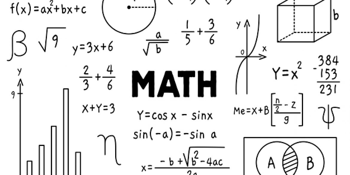
b-cubed – a^3 + b^3 = c^3
OnThere are many benefits of learning the game B-Cubed. It is fun, challenging, and requires you to use your math skills. Eventually, B-Cubed turns.
Read MoreThere are many benefits of learning the game B-Cubed. It is fun, challenging, and requires you to use your math skills. Eventually, B-Cubed turns.
Read MoreSpirit of Math is a global leader in after-school mathematics education end one of the best math website for tutors, teachers and other educators.
Read MoreThe Gauss Math Contest is an international math competition that is organized by the University of Waterloo in Canada. The competition is open to.
Read MoreMath isn’t a child’s favorite subject, but there are some ways to keep them engaged in learning. This program, which is a comprehensive approach.
Read MoreThe a cube minus b cube formula is a simple one and only requires three variables. It is expressed as a3 – b3 equation..
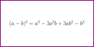
Read MoreThe a minus b cube whole equation is an integral form of acubes. It is a mathematical equation consisting of two terms, a and.
Read More